Tag Archives: physiotherapy martial arts

Managing myofascial pain

Myofascial pain is caused by a stimulus that sets off trigger points in your muscles. Triggers for myofascial pain can be factor that will affect the chemical balance of the body (stress and reduced sleep patterns). It will release some chemical substances in the body that can activate points of pain. This pattern is quite common with long term pain where normally the chemical balance is disturbed increasing the risk of long term pain.

Mechanical factors as long hours without movement in a static position can also increase muscle tightness and cause myofascial pain.

With myofascial pain, there are areas called trigger points. Normally, the patients report trigger points as “knots inside the muscle”. Trigger points are usually in the connective tissue (fascia) or in a tight muscle.

Myofascial pain syndrome is an ongoing or longer-lasting pain and can be managed with treatment.

Treatment includes aggravating factors management (stress management, improvement of sleep patterns);
Gentle stretching and respiratory exercises;
Soft tissue release;
Massage;
Acupuncture and dry needling;
Aerobic exercise;

New Concept of Tendinopathy: Scientific facts

See the source image

In the past few years research and new insights into tendon pathology has seen our understanding grow. Recently most therapists have had to stop themselves from saying the old term of “tendonitis” and learn the new term “tendinopathy”.

But it is not just the new term that has changed.

New studies suggest that under periods of excessive load a tendon will pass through the following continuum (Thanks to work of Dr Jill Cook and Craig Purdam). Their 2009 paper outlined a model that describes 3 different stages of tendinopathy reactive tendinopathy, tendon dysrepair and degenerative tendinopathy. It is helpful to think of these as a ‘continuum’ rather than 3 completely distinct phases:

Normal Tendon

↓ (reversible)

Reactive Tendinopathy

↓ (reversible)

Tendon Dysrepair (Failed Healing)

↓ (reversible)

Degenerative Tendinopathy

↓ (irreversible)

Rupture/Tear

Causes of Tendinopathy?

Tendon injuries are usually the result of increased loads and overuse. This leads to changes within the tendon, which make it harder for it to cope. The injuries may occur in the mid-portion or, more commonly, in the insertion. At both sites the pathological changes of the tendon appear to be the same. Despite the common pathological changes within the tendon, different treatment approaches are used specific to the site of the problem, and this has been shown to have better outcomes.

The slipped disc myth

Presentation2

The term ‘slipped disc’ is common when describing back pain, but it’s actually a bit of a misnomer. The discs in the spinal column can’t really slip out of place, but they can certainly suffer some damage (however, recent studies show that they can also be completely symptom free). Furthermore, one of the main factors of chronilow back pain is the lack of exercise and a poor life style which may affect the way a person is moving, posture and sleep patterns.

BASIC SPINE ANATOMY

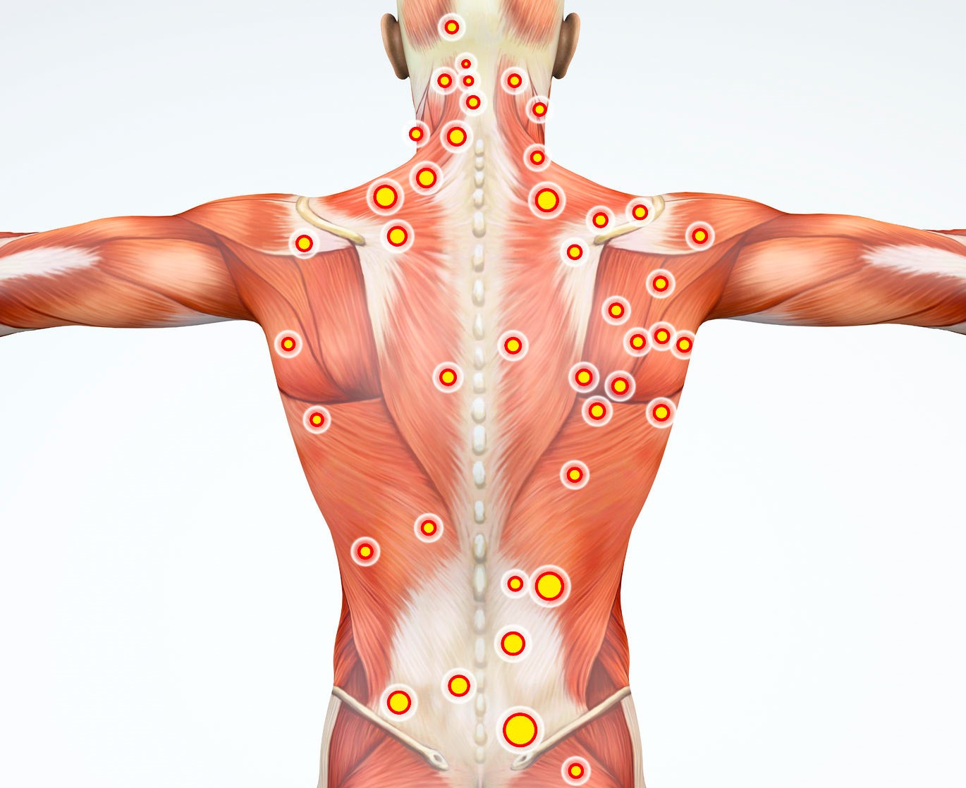
The spinal column is made up of 26 small bones called vertebrae. These bones link together to form a chain-like structure that allows flexibility in the torso but still protects the nerves connecting other parts of the body to the brain. In between the vertebrae is a small disc that cushions the spine by absorbing the shock from daily activities like walking and lifting.
These discs are made up of two parts. The outside is made of a tough, hard shell, while the inside (the nucleus pulposus) is soft and gel-like. Sometimes, the nucleus pulposus can protrude, or herniate, through the harder outer part of the disc, causing the disc to bulge and appear as if it has moved out of alignment with the spine. For this reason, the condition is colloquially referred to as a ‘slipped disc’ even though the disc itself has not really moved at all.
There are three kinds of disc herniation:
• Prolapse: Where the disc bulges out between vertebrae although its hard outer layer is still intact.
• Extrusion: Occurs where there is a tear in the hard outer layer of the disc which allows the nucleus pulposus to leak out. In these cases, the extruded tissue is still connected to the disc structure.
• Sequestration: The most extreme kind of herniation. This occurs when the nucleus pulposus completely escapes the disc structure and enters the spinal column.

CAUSES OF DISC HERNIATION

For most patients, disc herniation occurs as a normal part of the ageing process – the spine works very hard and sustains a good deal of wear and tear. As we get older, spinal discs lose some of their elasticity which leaves the more prone to cracking. Occasionally, injury to the spine or traumatic events (such as falling, repetitive activities or heavy lifting) can lead to herniation too.

SYMPTOMS OF HERNIATED DISCS

Herniated discs can cause compression on the nerves that run through the spinal column and this can be extremely painful. One of the most common symptoms of disc herniation in the lower part of the spine is sciatica.
Other symptoms can include pain, numbness, tingling and burning sensations. These symptoms can appear on one or both sides of the body, extending down the arms and the legs. Pain caused by herniated discs is often worse at night, or after extended periods of sitting or standing.

One of the most common things about herniated discs (suggested by recent studies) is that they can also be completely symptom free. Why? Pains is different from damage.

Note: If you are experiencing loss of bladder or bowel control in addition to back pain, this may indicate a more serious and rare problem called Cauda Equina syndrome. This condition is caused by the lower spinal nerve roots being compressed and requires urgent medical attention.

PHYSIOTHERAPY AND HERNIATED DISCS

As physiotherapists, we often hear patients talking about a ‘slipped disc’. Your discs are really strong tissues that sit between the bones of your spine. Because they are so strong, they simply cannot slip out of place, and neither can any other joints in your spine.

Most people who suffer from back pain do not need to have an MRI scan as part of their assessment. The majority of back pain complaints are caused by simple strains and sprains and a scan will not change the way patients are treated.

Physiotherapy can be very effective in relieving the pain and other symptoms associated with herniated discs. Techniques such as neural gliding, joint mobilisation and dry needling are all known to be effective in treating this condition. However, life style changes and improve exercising is the main answer suggested by recent studies.

Your physiotherapist will also show you movements and positions to help relieve the pain and symptoms of disc herniation, as well as giving you exercises to prevent further pain.

Spondylosis, spondylitis, spondylolysis and spondylolisthesis

Spondylosis, spondylitis, spondylolysis and spondylolisthesi are all different ways that the human spine can degenerate over time.

Spondylosis refers to degeneration of the spine. The term can be used to describe degeneration in the:

  • Neck – called cervical spondylosis
  • Lower back – called lumbar spondylosis
  • Middle back – called thoracic spondylosis

Two specific conditions may develop from this degeneration – degenerative disc disease and spinal osteoarthritis – depending on if the degeneration occurs in the discs or in the facet joints, respectively. The development of bone spurs may also occur as part of the degeneration of the spine with spondylosis and with either of the degenerative conditions.

Spondylolysis most of the vertebra consist of a body, pedicle, lamina, pars interarticularis, transverse process, spinous process and superior articular facets. Together these parts form joints that link the vertebrae together. The pars interarticularis is the weakest link in the chain and spondylolysis is basically a stress fracture of the pars interarticularis (Scotty dog).

Spondylitis Tropheryma whipplei.jpg

Spondylitis is an inflammation of the vertebra. It is a form of spondylopathy. In many cases spondylitis involves one or more vertebral joints as well, which itself is called spondylarthritis

Spondylolisthesis occurs when a bone from the lower spine (a vertebra) slips out of position.

This is not to be confused with a slipped disc, when one of the spinal discs inbetween the vertebrae ruptures.

All of these issues can be asymptomatic and never cause pain and discomfort. They can also lead to minor tightening or stiffening, and in sever instances can result in intense chronic pain that leads to a loss of quality of life.

Improving our posture and core tone will go a long way to supporting the spine through life. Having a spine that suffers with this conditions is not the end of the world but this type of spine requires more awareness and muscular support to get through life.

Acute muscle soreness: How to treat it?

Acute muscle soreness is the pain felt in muscles during and immediately after strenuous physical exercise.  The pain appears within a minute of contracting the muscle and disappears within two or three minutes or up to several hours after relaxing it.

The following causes have been proposed for acute muscle soreness:

  • Accumulation of chemical end products of exercise in muscle cells, such as H+
  • Tissue edema caused by the shifting of blood plasma into the muscle tissue during contraction
  • Muscle fatigue (the muscle tires and cannot contract any more)

Acute muscle soreness is one form of exercise-induced muscle damage, the other being delayed onset muscle soreness, which appears between 24 and 72 hours after exercise.

Delayed onset muscle soreness (DOMS). How to treat it?

delayed muscle pain

Find us on Facebook and leave your like.

Delayed onset muscle soreness (DOMS), also called muscle fever, is the pain and stiffness felt in muscles several hours to days after unaccustomed or strenuous exercise.

The soreness is felt most strongly 24 to 72 hours after the exercise. It is thought to be caused by eccentric (lengthening) exercise, which causes microtrauma to the muscle fibers. After such exercise, the muscle adapts rapidly to prevent muscle damage, and thereby soreness, if the exercise is repeated.

Delayed onset muscle soreness is one symptom of exercise-induced muscle damage. The other is acute muscle soreness, which appears during and immediately after exercise.

The soreness is perceived as a dull, aching pain in the affected muscle, often combined with tenderness and stiffness. The pain is typically felt only when the muscle is stretched, contracted or put under pressure, not when it is at rest. This tenderness, a characteristic symptom of DOMS, is also referred to as “muscular mechanical hyperalgesia”.

Although there is variance among exercises and individuals, the soreness usually increases in intensity in the first 24 hours after exercise. It peaks from 24 to 72 hours, then subsides and disappears up to seven days after exercise.

The mechanism of delayed onset muscle soreness is not completely understood, but the pain is ultimately thought to be a result of microtrauma – mechanical damage at a very small scale – to the muscles being exercised.

Delayed onset muscle soreness is thought to be a result of microscopic tearing of the muscle fibers. The amount of tearing (and soreness) depends on how hard and how long you exercise and what type of exercise you do. Any movement you aren’t used to can lead to DOMS, but eccentric muscle contractions (movements that cause muscle to forcefully contract while it lengthens) seem to cause the most soreness. Examples of eccentric muscle contractions include going down stairs, running downhill, lowering weights and the downward motion of squats and push-ups. In addition to small muscle tears there can be associated swelling in a muscle which may contribute to soreness.

Treatment:

If you do find yourself sore after a tough workout or competition, try these methods to deal with your discomfort. Although not all are backed up with research, many athletes report success with some of the following methods:

Active recovery: Performing easy low-impact aerobic exercise increasing blood flow and is linked with diminished muscle soreness. After an intense workout or competition, use this technique as a part of your cool down;

Rest: If you simply wait it out, soreness will go away in 3 to 7 days with no special treatment.

Contrast water therapy and Ice immersion therapy: Of the studies that have looked at the effects of ice baths, cold water immersion and contrast water therapy on exercise recovery and muscle soreness, most offer inconclusive or contradictory findings.

Ankle injuries and anatomy of the ankle

anklelock foot lock injuries

Find us on Facebook and leave your like.

The ankle, or the talocrural region, is the region where the foot and the leg meet. The ankle includes three joints:

1-the ankle joint proper or talocrural joint;

2- the subtalar joint;

3-the tibiofibular joint.

The movements produced at this joint are dorsiflexion and plantarflexion of the foot. In common usage, the term ankle refers exclusively to the ankle region. In physio-medical terminology, “ankle” (without qualifiers) can refer broadly to the region or specifically to the talocrural joint.

The bony architecture of the ankle consists of three bones: the tibia, the fibula, and the talus. The ankle joint is bound by the strong deltoid ligament and three lateral ligaments: the anterior talofibular ligament, the posterior talofibular ligament, and the calcaneofibular ligament. We can not forget to mention the syndesmotic ligament. This ligament spans the syndesmosis, which is the term for the articulation between the medial aspect of the distal fibula and the lateral aspect of the distal tibia. An isolated injury to this ligament is often called a high ankle sprain.

The bony architecture of the ankle joint is most stable in dorsiflexion. Thus, a sprained ankle is more likely to occur when the ankle is plantar-flexed, as ligamentous support is more important in this position. The classic ankle sprain involves the anterior talofibular ligament (ATFL), which is also the most commonly injured ligament during inversion sprains. Another ligament that can be injured in a severe ankle sprain is the calcaneofibular ligament.

Risk Factors  

Factors that increase your chance of getting an ankle sprain include:

  • Playing sports especially with the wrong type of shoe
  • Sports as sambo, MMA, judo where the ankle is a target itself
  • Walking on uneven surfaces
  • Weak ankles from a previous sprain
  • Having:
    • Poor coordination
    • Poor balance
    • Poor muscle strength and tight ligaments
    • Loose joints
  • Certain footwear (for example high heels)
Symptoms  

Symptoms of an ankle sprain may include:

  • Pain, swelling, and bruising around the ankle
  • Worsening of pain when walking, standing, pressing on the sore area, or moving the ankle inward
  • An inability to move the ankle joint without pain
  • A popping or tearing sound at the time of the injury (possibly)
Diagnosis

An ankle sprain may not require a visit to the doctor. However, you should call your  physiotherapist or doctor if you have any of the following:

  • Inability to move the ankle without significant pain
  • Inability to put any weight on that foot
  • Significant swelling or bruising
  • Pain over a bony part of your foot or ankle
  • Pain that interferes significantly with walking
  • Pain not relieved by ice, pain relief medication, and elevation
  • Numbness in the leg, foot, or ankle
  • Pain that does not improve in 5-7 days
  • Uncertainty about the severity of the injury
  • Uncertainty about how to care for this injury

The physiotherapist or doctor will ask about your symptoms and how your injury occurred. An examination of your ankle will be done to assess the injury.

Images may need to be taken of your ankle. This can be done with:

  • X-rays
  • MRI

Ankle sprains are graded according to the damage to the ligaments. The more ligaments involved, the more severe the injury.

Grade 1
  • Some minor tearing of ligament tissue
  • Ankle remains stable
Grade 2
  • Partial tearing of ligament tissue
  • Mild instability of the joint
  • Usually involves damage to 2 ankle ligaments
Grade 3
  • Complete tearing of 2 or 3 of the ligaments
  • Significant instability of the joint
Treatment  

Most sprains heal well. Treatment for a sprained ankle includes:

  • Rest—Avoid putting any pressure on your ankle by not walking on it. Using crutches will let you bear partial weight. This is allowed early on, except when all three ligaments are torn.
  • Ice—Apply ice or a cold pack to the ankle for 15-20 minutes, 4 times a day for at least 2-3 days. This helps reduce pain and swelling. Wrap the ice or cold pack in a towel. Do not apply the ice directly to your skin.
  • Compression—Wrap your ankle in an elastic compression bandage. Wrap from the toes going up toward the knee. This will limit swelling of your ankle and foot.
  • Elevation—Keep your ankle raised above the level of your heart as much as you can for 48 hours. This will help drain fluid and reduce swelling.
  • Oral pain medicine such as, ibuprofen, naproxen, acetaminophen, aspirin or topical pain medicines, such as creams and patches that are applied to the skin
  • Physiotherapy—Begin exercises to restore flexibility, balance, range of motion, and strength of the muscles around your ankle as recommended by your physiotherapist or doctor. You may benefit from working with a physiotherapist that can teach you the exercises and make sure that you are performing them correctly. Physiotherapists can use different techniques such as mobilization, manipulation, taping, proprioception training, etc. This kind of treament makes the difference to increase your rate of recovery.
  • Brace—You may need to wear a brace or walking boot to prevent your ankle from moving. In many cases, a brace, which stabilizes and compresses the ankle, will allow for early weight bearing and an earlier return to activity. You will be rehabilitating the ankle as it heals. If you play sports, you may need to wear an ankle brace or tape your ankle when you return to play.
  • Leg cast—If you have a severe sprain, your doctor may recommend a short leg cast for 2-3 weeks, but this is very rare. In many cases, there are special braces that can be used instead of a cast.
  • Surgery—Surgery is rarely needed to repair an ankle sprain. Yes, physiotherapy is the best option. However, it may be necessary to repair a third degree sprain in which all three ligaments are torn.

I hurt my elbow practicing jiu jitsu. Any idea what happened?

Diapositiva1

The elbow joint is a complex hinge joint formed between the distal end of the humerus in the upper arm and the proximal ends of the ulna and radius in the forearm. The elbow allows for the flexion and extension of the forearm relative to the upper arm, as well as rotation of the forearm and wrist.

An extensive network of ligaments surrounding the joint capsule helps the elbow joint maintain its stability and resist mechanical stresses. The radial and ulnar collateral ligaments connect and maintain the position of the radius and ulna relative to the epicondyles of the humerus. The annular ligament of the elbow extends from the ulna around the head of the radius to hold the bones of the lower arm together. These ligaments allow for movement and stretching of the elbow while resisting dislocation of the bones.

Because so many muscles originate or insert near the elbow, it is a common site for injury. One common injury is lateral epicondylitis, also known as tennis elbow, which is an inflammation surrounding the lateral epicondyle of the humerus. Six muscles that control backward movement (extension) of the hand and fingers originate on the lateral epicondyle. Repeated strenuous striking while the muscles are contracted and against force – such as that occurring with the backhand stroke in tennis – causes strain on the tendinous muscle attachments and can produce pain around the epicondyle. Rest for these muscles will usually bring about recovery.

One of the most common injuries in jiujitsu, judo, wrestling, boxing and muay thai is an elbow sprain. An elbow sprain is stretching or tearing of the ligaments that stabilize the elbow. Ligaments are strong bands of tissue that cross joints and connect bones to each other. The sprain occurs when  the joint is push beyond its normal range of motion. E.g. during armbars.

However, there are several injuries that can affect the elbow:  muscle strain, tendon tear, ligament sprain, hair line fracture, slight dislocation, etc.  Often the ligament is affected. A skilled physiotherapist can help you regarding these common questions during the assessment.

How could you manage that?

For the first 48-72 hours think of:

  • Paying the PRICE – Protect, Rest, Ice, Compression, Elevation; and
  • Do no HARM – no Heat, Alcohol, Running or Massage.
  • Physiotherapy after 72 hours.

The aim of physiotherapy is to help restore movement and normal body function in cases of illness, injury and disability. Physiotherapists take a holistic approach, looking at the body as a whole rather than focusing on the individual factors of an injury or illness.

Physiotherapists use a wide range of treatment techniques and approaches. Some of these are described below:

Movement and exercise, taping, acupuncture, manual therapy, dry needling, hydrotherapy, etc.


One of the most common injuries in jujitsu is “joint hyper extension”. This occurs when a joint, usually an elbow, is push beyond its normal range of motion. – See more at: http://submissions101.com/articles/jiu-jitsu-joint-injuries-and-repair.html#sthash.QG7ruoha.dpuf

What is the treatment for a knee ligament injury?

knee brazilian jiu jitsu

Self-help treatment

For the first 48-72 hours think of:

  • Paying the PRICE – Protect, Rest, Ice, Compression, Elevation; and
  • Do no HARM – no Heat, Alcohol, Running or Massage.

Paying the PRICE:

  • rotect your injured knee from further injury.
  • est your affected knee for 48-72 hours following injury. Consider the use of crutches to keep the weight off your injured knee. However, many doctors say that you should actually not keep your injured knee immobile for too long. You can usually start some exercises to help keep your knee joint moving and mobile. Start these as soon as you can tolerate the exercises without them causing too much pain. You can ask your doctor when you can start to move your knee joint and what exercises you should do.
  • ce should be applied as soon as possible after your knee injury – for 10-30 minutes. Less than 10 minutes has little effect. More than 30 minutes may damage the skin. Make an ice pack by wrapping ice cubes in a plastic bag or towel. (Do not put ice directly next to skin, as it may cause ice-burn.) A bag of frozen peas is an alternative. Gently press the ice pack on to your injured knee. The cold from the ice is thought to reduce blood flow to the damaged ligament. This may limit pain and inflammation. After the first application, some doctors recommend reapplying for 15 minutes every two hours (during daytime) for the first 48-72 hours. Do not leave ice on while asleep.
  • ompression with a bandage will limit swelling, and help to rest your knee joint. A tubular compression bandage can be used. Mild pressure that is not uncomfortable or too tight, and does not stop blood flow, is ideal. A pharmacist will advise on the correct size. Remove before going to sleep. You may be advised to remove the bandage for good after 48 hours. This is because the bandage may limit movement of the joint which should normally be moving more freely after this time. However, bandages of the knee are sometimes kept on for longer to help keep swelling down and to keep the affected knee more comfortable. Ask your doctor what is best in your case.
  • levation aims to limit and reduce any swelling. For example, keep your foot on the affected side up on a chair when you are sitting. It may be easier to lie on a sofa and to put your foot on some cushions. When you are in bed, put your foot on a pillow. The aim is that your affected knee should be above the level of your heart.
  • Anti-inflammatory painkillers. If you need to take these for more that three days, then you will need to discuss this with your doctor or pharmacist.

Avoid HARM for 72 hours after injury. That is, avoid:

  • eat – for example, hot baths, saunas, heat packs. Heat has the opposite effect to ice on the blood flow. That is, it encourages blood flow. So, heat should be avoided when inflammation is developing. However, after about 72 hours, no further inflammation is likely to develop and heat may then be soothing.
  • lcoholic drinks, which can increase bleeding and swelling and decrease healing.
  • unning or any other form of exercise which may cause further damage.
  • assage, which may increase bleeding and swelling. However, as with heat, after about 72 hours, gentle massage may be soothing.

Physiotherapy

(Important: Massage therapists are not physiotherapists. Physiotherapists must be HPC and CSP registered. Once registered, physiotherapists must continue to meet the standards of proficiency that are relevant to their scope of practice).

This may be helpful after some knee ligament injuries. Physiotherapy may help to improve the range of movement in your injured knee. Exercises may also be suggested to help strengthen the muscles that support your knee joint. If you are considering surgery to repair a torn knee ligament, you may be advised to have physiotherapy before the operation.

The Physiotherapists will first take a detailed history of your condition together with any relevant past medical history.

A physical assessment will then be undertaken to determine the clinical diagnosis of your problem. The physiotherapist will discuss with you a proposed treatment plan and will give you an estimate as to the length of treatment required and the proposed outcome of your treatment.

The initial assessment will probably last up to one hour and subsequent treatment sessions will last between thirty and forty-five minutes approximately.

Every opportunity will be given to you to ask questions about your condition and advice regarding its management and prevention in the future.